On April 17, 2024, the U.S. Food and Drug Administration (FDA) has approved the optical imaging agent Lumisight (pegulicianine, LUM015) and the Lumicell Direct Visualization System (DVS) - collectively referred to as LumiSystemTM for use in fluorescence imaging in adult patients with breast cancer as an adjunct for the intraoperative detection of cancerous tissue within the resection cavity after the removal of the primary specimen during lumpectomy surgery.
Breast-conserving surgery
Breast cancer is the most commonly occurring cancer in women and the most common cancer overall. The main types of treatment for breast cancer are surgery, radiation therapy (RT), chemotherapy (CT), endocrine (hormone) therapy (ET), and targeted therapy.
Breast-conserving surgery (sometimes called a lumpectomy) is a primary treatment method for non-invasive and early-stage breast cancer. In this procedure, the surgeon removes the tumor along with some surrounding tissue, which is then sent to a pathologist for postoperative evaluation. However, traditional pathological assessment is a time-consuming process. If the pathologist finds cancer cells at the margins of the removed tissue (positive margins), additional surgery may be required to ensure all cancer cells are completely removed from the patient.
Worryingly, studies indicate that even when pathological assessment shows negative margins, about 14% of patients still have residual cancer cells. This means that no current surgical method can completely address this issue.
Lumicell Direct Visualization System (DVS)
To meet these clinical needs, Lumicell has developed a new imaging system composed of a proprietary optical imaging agent, a handheld imaging device, and patient-calibrated tumor detection software. Together, these components form the Lumicell Direct Visualization System (DVS), which enables surgeons to scan the breast cavity post-lumpectomy, in real-time, to detect and resect residual cancer that may have otherwise been missed, potentially sparing some patients from second surgeries.
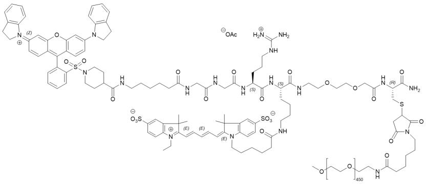
Lumisight (pegulicianine), a fluorescent imaging drug, consists of a commercially available fluorescence quencher molecule (QSY21) attached through a GGRK peptide to a 20-kD polyethylene glycol (PEG) and a Cy5 fluorophore.

Figure 1. Structure of pegulicianine, source: wikipedia.com
Pegulicianine is optically inactive when intact and produces a fluorescent signal after its peptide chain is cleaved by cathepsins and matrix metalloproteases (MMPs). The levels of these enzymes are higher in and around tumor and tumor-associated cells than normal cells. This enzymatic cleavage results in “fragment 2” and “fragment 3”, which are optically active metabolites that emit fluorescence, as well as “fragment 1” containing the fluorescence quencher that keeps the intact molecule optically inactive.
This approval is based on a pivotal Phase 3 trial named INSITE (NCT03686215). The study enrolled 406 patients with primary invasive breast cancer and/or ductal carcinoma in situ (DCIS) from 14 hospitals in the United States.
Among the 357 patients randomized to the treatment arm, 27 (7.6%; 95% CI [5.0%, 10.8%]) had residual tumors removed from the margins guided by the Lumicell DVS after standard lumpectomy, including 22 from cavity orientations deemed negative on standard margin evaluation. By excising margins guided by the Lumicell DVS, 9 out of 62 patients (14.5%) with initially positive margins were converted to final negative margins, potentially avoiding a second surgery in these patients. In the analysis of residual cancer margins, the Lumicell DVS demonstrated a specificity of 85.2% (95% CI [83.7%, 86.6%]) and a sensitivity of 49.3% (95% CI [37.0%, 61.6%]).
LUMISIGHT was generally well-tolerated with a serious adverse event rate of 0.5%, similar to that of other contrast agents.
Conclusion
The approval of pegulicianine and the Lumicell Direct Visualization System (DVS) provides more accurate and real-time imaging guidance for breast cancer surgery. This advancement is expected to reduce the risk of residual cancerous tissue post-surgery, thereby decreasing the need for additional surgeries. With this innovative technology, patients can more quickly understand the outcomes of their surgeries, avoid unnecessary pain and costs, and experience improved treatment efficiency and overall satisfaction.
Biopharma PEG, a top PEG supplier, offers high-purity PEG linkers that enhance water solubility. This enables efficient biolabeling in imaging and diagnostic research and development, making it easier to conduct experiments and studies in this field.
References:
[1] https://www.fda.gov/drugs/news-events-human-drugs/fda-approves-imaging-drug-assist-detection-cancerous-tissue-following-lumpectomy
[2] Whitley MJ, Cardona DM, Lazarides AL, Spasojevic I, Ferrer JM, Cahill J, Lee CL, Snuderl M, Blazer DG 3rd, Hwang ES, Greenup RA, Mosca PJ, Mito JK, Cuneo KC, Larrier NA, O'Reilly EK, Riedel RF, Eward WC, Strasfeld DB, Fukumura D, Jain RK, Lee WD, Griffith LG, Bawendi MG, Kirsch DG, Brigman BE. A mouse-human phase 1 co-clinical trial of a protease-activated fluorescent probe for imaging cancer. Sci Transl Med. 2016 Jan 6;8(320):320ra4. doi: 10.1126/scitranslmed.aad0293. PMID: 26738797; PMCID: PMC4794335.

